
Glaucoma
Glaucoma can cause blindness if it is left untreated. Only about half of the estimated three million Americans who have glaucoma are even aware that they have the condition. When glaucoma develops, patients usually don’t have any early symptoms as the disease progresses slowly. In this way, glaucoma can steal your sight very gradually. Fortunately, early detection and treatment with glaucoma eyedrops, glaucoma lasers or both can help preserve your vision.
For Appointments: Call (516) 794-2020

What is Glaucoma?
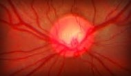
Glaucoma is a disease that damages the eye’s optic nerve. The optic nerve is connected to the retina — a layer of light-sensitive tissue lining the back of the eye — and is made up of many nerve fibers, like an electric cable is made up of many wires. It is the optic nerve that sends signals from your retina to your brain, where these signals are interpreted as the images you see.
In the healthy eye, a clear fluid called aqueous (pronounced AY-kwee-us) humor circulates inside the front portion of your eye. To maintain a constant healthy eye pressure, your eye continually produces a small amount of aqueous humor while an equal amount of this fluid flows out of your eye. If you have glaucoma, the aqueous humor does not flow out of the eye properly. Fluid pressure in the eye builds up and, over time, causes damage to the optic nerve fibers.
If you have glaucoma, it is important to tell your ophthalmologist about your other medical conditions and all other medications you currently take. Bring a list of your medications with you to your eye appointment. Also tell your primary care doctor and any other doctors caring for you what glaucoma medication you take.
Endoscopic CycloPhotocoagulation (ECP)
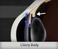
Endoscopic CycloPhotocoagulation, or ECP, is an exciting development in the management of many types of glaucoma including the more common open-angle glacuoma and narrow-angle glaucoma. ECP is performed on an outpatient basis. In this procedure, the ciliary body of the eye, which creates fluid, is treated with a laser. This reduces fluid production that, in turn, reduces intra-ocular pressure. The ciliary body is a small gland running around the circumference of the eye located behind the iris.
Endoscopic CycloPhotocoagulation is often performed on patients at the time of cataract surgery. It can also be performed on those patients who had SLT or ALT laser procedures, glaucoma filtration surgery or other surgical procedures that were not successful at controlling intra-ocular pressure.

ECP allows the surgeon to view the area through an endoscopic camera, which aids in the very precise placement of the laser beam used for treatment.
ECP has proven to be an effective way to reduce intra-ocular pressure. Studies have shown that the majority of patients have their glaucoma medications reduced or completely eliminated after the procedure and are no longer at risk of loss of vision from glaucoma.
Endoscopic CycloPhotocoagulation is for those who:
- have glaucoma or higher than normal intra-ocular pressure, including those with cataracts
- have not responded well to medications, laser, or other surgical treatments.
What to expect on procedure day:
ECP is most often performed at the same time as cataract surgery, after the cataract has been removed from the eye. No additional incisions are required. The ECP probe uses tiny, optical fibers to illuminate, view and treat the ciliary body with laser energy. Approximately 20 to 40 laser applications will be administered.
Your eye pressure will be checked shortly after your procedure and drops may be prescribed to alleviate any soreness or swelling inside your eye. You may also receive oral medications to control eye pressure or inflammation.You should relax for the rest of the day. Follow-up visits are necessary to monitor eye pressure. It may take a few weeks to see the full pressure-lowering effect of this procedure. Most patients resume normal activities within a few days.
Realistic expectations:
The effect of the surgery may wear off over time, but the majority of patients have their pressure lowered and many can reduce or eliminate their need for glaucoma medications. However, this procedure and other glaucoma surgical procedures do not restore lost vision. Serious complications with Endoscopic CycloPhotocoagulation are rare, but like any surgical procedure, ECP may have some risks. Prior to your procedure, you will be given additional information that will allow you to make an informed decision. At Berke Eye Care, you can be sure that all your questions are answered to your satisfaction.
If you would like more information about this procedure , you can make an appointment or contact Berke Eye Care at 516-794-2020.
Glaucoma surgery
In some patients with glaucoma, surgery is recommended. Glaucoma surgery improves the flow of fluid out of the eye, resulting in lower eye pressure.
Laser trabeculoplasty
A surgery called laser trabeculoplasty is often used to treat open-angle glaucoma. There are two types of trabeculoplasty surgery: argon laser trabeculoplasty (ALT) and selective laser trabeculoplasty (SLT).
During ALT surgery, a laser makes tiny, evenly spaced burns in the trabecular meshwork. The laser does not create new drainage holes, but rather stimulates the drain to function more efficiently.
With SLT, a low level energy laser targets specific cells in the mesh-like drainage channels using very short applications of light. The treatment has been shown to lower eye pressure at rates comparable to ALT.
Even if laser trabeculoplasty is successful, most patients continue taking glaucoma medications after surgery. For many, this surgery is not a permanent solution. Nearly half who receive this surgery develop increased eye pressure again within five years. Many people who have had a successful laser trabeculoplasty have a repeat treatment.
Laser trabeculoplasty can also be used as a first line of treatment for patients who prefer not to use glaucoma eyedrops every day.
Laser iridotomy
Laser iridotomy is recommended for treating people with closed-angle glaucoma and those with very narrow drainage angles. A laser creates a small hole about the size of a pinhead through the iris to improve the flow of aqueous fluid to the drainage angle. This hole is so small that it is not visible without magnification.
Peripheral iridectomy
When laser iridotomy is unable to stop an acute closed-angle glaucoma attack, or is not possible for other reasons, a peripheral iridectomy may be performed. Performed in an operating room, a small piece of the iris is removed, giving the aqueous fluid access to the drainage angle again. Because most cases of closed-angle glaucoma can be treated with glaucoma medications and laser iridotomy, peripheral iridectomy is rarely necessary.
Trabeculectomy
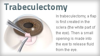
In trabeculectomy, a small flap is made in the sclera (the outer white coating of your eye). A filtration bleb, or reservoir, is created under the conjunctiva — the thin, filmy membrane that covers the white part of your eye. Once created, the bleb looks like a bump or blister on the white part of the eye above the iris, but the upper eyelid usually covers it. The aqueous humor can now drain through the flap made in the sclera and collect in the bleb, where the fluid will be absorbed into blood vessels around the eye.
Eye pressure is effectively controlled in three out of four people who have trabeculectomy. Although regular follow-up visits with your doctor are still necessary, many patients no longer need to use eyedrops. If the new drainage channel closes or too much fluid begins to drain from the eye, additional surgery may be needed.
Glaucoma Shunt
How does the Ex-PRESS mini shunt work?
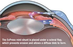
The Ex-PRESS mini shunt is a relatively new device, available in the USA since 2002. Over 12,000 implantations have been performed worldwide. It is a straightforward procedure, in which you divert the fluid through an extremely small tube to the outside of the eye. You neither cut sclera, nor iris. Although the device is as small as a grain of rice, it acts just like a heart stent keeping a pathway open so blood (or fluid) can successfully go around the blockage. The Ex-PRESS provides precise control of the amount of fluid that is allowed to flow out, helping the eye maintain a healthy level of internal pressure.
The Ex-PRESS mini glaucoma shunt provides effective long term control of intraocular pressure, with a success rate of about 94% . It can be done following cataract surgery. The Ex-PRESS has an equally effective intraocular pressure control compared with trabeculectomy, but is safer than trabeculectomy in the short term. In most cases, patients virtually eliminate their need for glaucoma eye drops after Ex-PRESS surgery.
Aqueous shunt surgery
If trabeculectomy cannot be performed, aqueous shunt surgery is usually successful in lowering eye pressure.
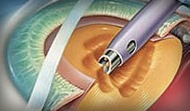
An aqueous shunt is a small plastic tube or valve connected on one end to a reservoir (a roundish or oval plate). The shunt is an artificial drainage device and is implanted in the eye through a tiny incision. The shunt redirects aqueous humor to an area beneath the conjunctiva (the thin membrane that covers the white part of your eye). The fluid is then absorbed into the blood vessels. When healed, the reservoir is not easily seen unless you look downward and lift your eyelid.
Important things to remember about glaucoma:
There are a number of ways to treat glaucoma. While some people may experience side effects from glaucoma medications or glaucoma surgery, the risks of side effects should always be balanced with the greater risk of leaving glaucoma untreated and losing vision.
If you have glaucoma, preserving your vision requires strong teamwork between you and your doctor. Your doctor can prescribe treatment, but it’s important to do your part by following your treatment plan closely. Be sure to take your medications as prescribed and see your ophthalmologist regularly.
Other Services

Cataract
If your vision has become blurry, cloudy or dim, or things you see are not as bright or colorful as they used to be, a cataract may have developed in one or both of your eyes.

Routine Eye Exams
Routine eye exam's are a very important part of keeping your eyes healthy. Most eye exam's are simple and comfortable and do not take more than 45 to 90 minutes.

Allergy Testing
At Berke Eye Care, we now offer in house allergy testing. With over 200+ allergens out there,
it is great first step to effective allergy treatment and is usually covered by most insurance plans.
Better Eye Care is Our Mission
CALL US. Same Day Appointments are Available.
Ophthalmology in Long Island